Fig. 4.
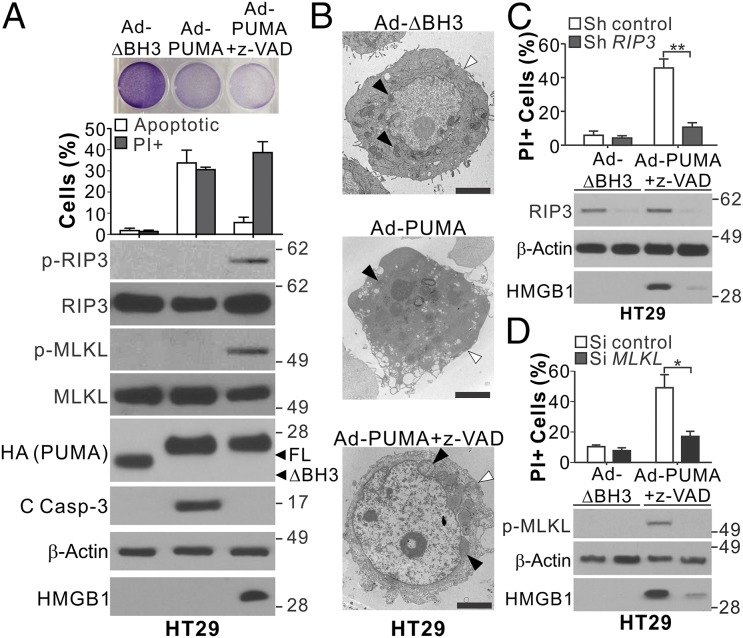
PUMA expression alone can induce necroptosis and RIP3 and MLKL phosphorylation in RIP3-expressing cells with caspase inhibition. (A) HT29 cells with or without pretreatment with 10 μM z-VAD were infected with control (BH3 domain deleted; ΔBH3) or PUMA-expressing adenovirus (Ad-PUMA). (Upper) Crystal violet staining at 24 h. (Middle) analysis of apoptosis and PI staining at 48 h. (Lower) Western blots of indicated proteins and HMGB1 release at 24 h. (B) Representative TEM pictures of HT29 cells treated as in A for 24 h. Black arrowheads indicate mitochondria, and white arrowheads indicate plasma membranes. (Scale bars: 2 μm.) (C) HT29 cells stably transfected with control or RIP3 shRNA were treated and analyzed as in A. (D) HT29 cells transfected with control scrambled or MLKL siRNA were treated and analyzed as in A. Values in A, C, and D are expressed as mean ± SD. n = 3. *P < 0.05; **P < 0.01.

