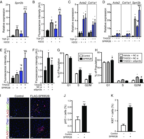
Fig. 2.
Synergistic activation of Sprr2b by TGF-β1 and H2O2 is required for CF proliferation. (A–C) Primary neonatal CFs were treated with combinations of TGF-β1 (10 ng/mL) and H2O2 (50 μM) for 24 h. (A) qRT-PCR was performed to evaluate Sprr2b expression (n = 6). One-way ANOVA. (B) CyQuant dye DNA incorporation was performed to evaluate proliferation (n = 8). One-way ANOVA. (C) qRT-PCR for Acta2 and Col1a1 was performed to evaluate myofibroblast activation (n = 6). Two-way ANOVA with Bonferroni post hoc. (D) SPRR2B transfection of CFs followed by qRT-PCR for Acta2 and Col1a1 (n = 6). Two-way ANOVA with Bonferroni post hoc. (E) CyQuant assay was used to evaluate the impact of SPRR2B overexpression on CF proliferation (n = 8). One-way ANOVA. (F) CyQuant assay was used to evaluate the effect of siRNA-mediated Sprr2b knockdown on TGF-β1/H2O2–induced CF proliferation (n = 8). One-way ANOVA. NC, no-target control siRNA. (G) Flow cytometry shows G2/M and S populations increase in response to SPRR2B overexpression in CFs relative to control (n = 3). One-way ANOVA. (H) Flow cytometry reveals siRNA-mediated knockdown of Sprr2b in CFs blocks the increase in G2/M and S populations by TGF-β1/H2O2 treatment, relative to control (n = 3). One-way ANOVA. (I) Primary CFs were transfected with control or Flag-SPRR2B expression vectors (transfection efficiency was ∼36%). Immunofluorescent detection of FLAG and phosphohistone H3 or Ki67 reveals increased mitosis and proliferation in SPRR2B-transfected CFs. (Scale bar: 50 μm.) (J) Quantification of pH3+ cells from I. (K) Quantification of Ki67+ cells from I. n = 6 wells with five nonoverlapping fields of view each. Unpaired Student’s t test. qRT-PCR data normalized to Gapdh. All data represent mean ± SEM. *P < 0.05, **P < 0.01, ***P < 0.001, ****P < 0.0001 compared with control. #P < 0.05 compared with second condition; &P < 0.05 compared with third condition.

