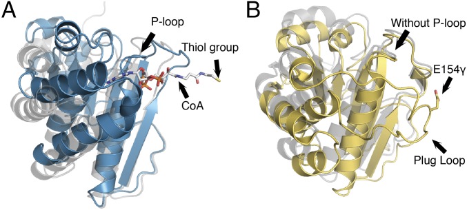
Fig. 6.
Domain III comparison for MtPFOR and MtOOR. (A) Domain III of MtPFOR with CoA bound (teal) and without CoA bound (gray). In PFOR, domain III anchors pyrophosphate and adenine of CoA and swings toward the active site, initiating the second electron transfer step in the PFOR reaction. (B) Domain III of MtOOR swung-in toward the active site [yellow; PDB ID code 5C4I (12)] and swung-out away from the active site [gray; PDB ID code 5EXE (13)]. In OOR, domain III contains a plug loop housing Glu154γ. The plug-loop movement facilitates oxalate decarboxylation. 19γARGVVM24γ in OOR spatially corresponds to the location of the P-loop in PFOR, but the motif adapts a different geometry that does not have sufficient space to bind a phosphate.

