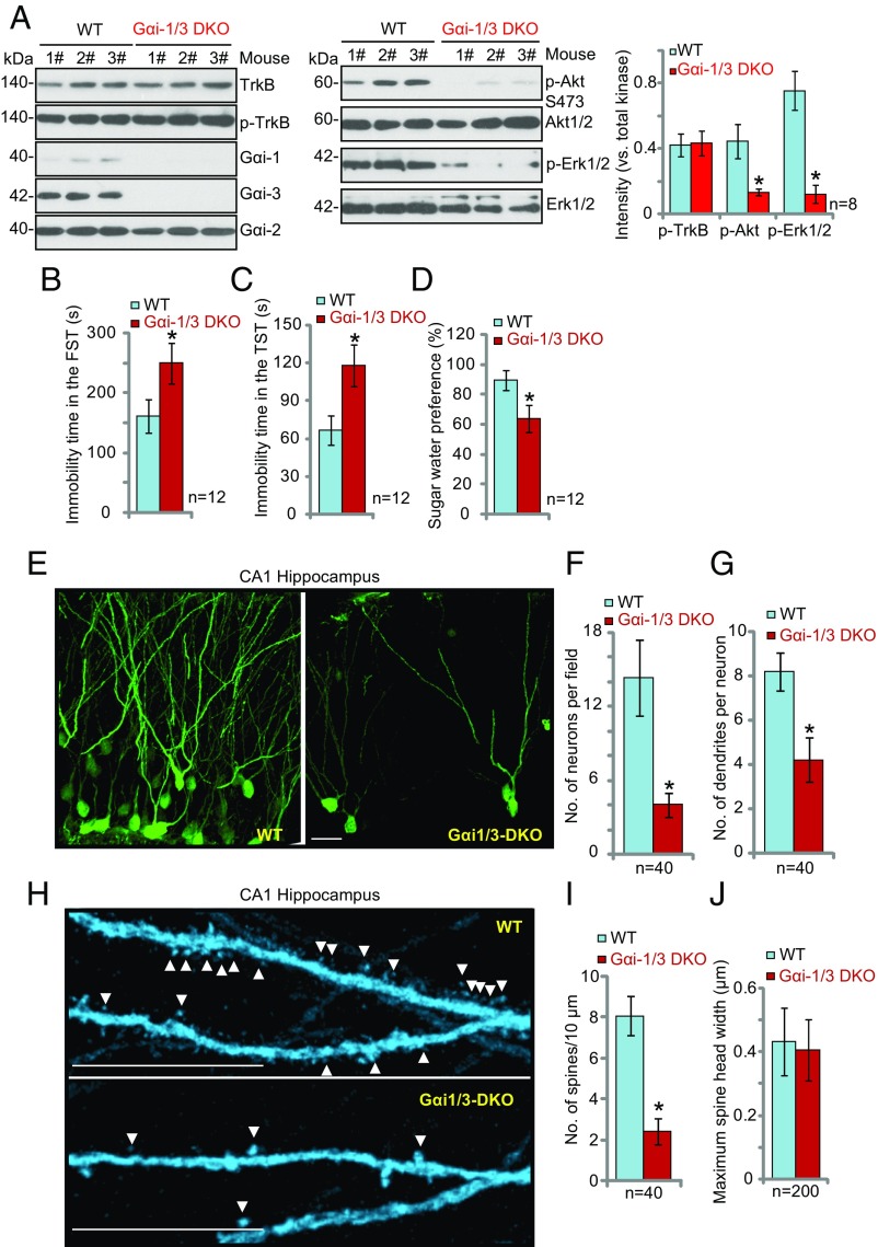
Fig. 7.
Severe depressive-like behaviors in Gαi1/3-DKO mice. (A) Depletion of Gαi1 and Gαi3 in the DKO mice disrupts signaling. The expression of the listed proteins in the CA1 hippocampus of WT and Gαi1/3-DKO mice was examined by Western blot analysis. (B–D) DKO mice display depressive behaviors. For both WT and Gαi1/3-DKO mice, the FST (B), TST (C), and sucrose water preference test (D) were performed. (E–J) Analysis of DKO hippocampal CA1 neuronal morphology. (E and H) Representative images of CA1 pyramidal hippocampal neuronal morphology. Arrowheads indicate spines. (Scale bars: 25 μm in E and 5 μm in H.) (F) The number of neurons in randomly selected 200 × 200 μm fields was counted. (G and I) The number of secondary dendrites (G) and spines (I) in 40 random neurons were counted. Spines were analyzed from 30-μm-long apical dendritic segments (50–80 μm from soma). (J) The maximum spine width of 200 spines from 10 randomly selected neurons was measured by Image J software. *P < 0.001 vs. WT mice.

