
Figure 3. Sequential activation of NF-κB and STAT3 is required for HER2-mediated expansion of cancer stem-like cells.
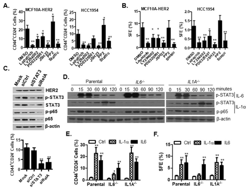
A–B. Cells were treated with lapatinib (2μM), LY294002 (10μM), PD0325901 (100nM), JSH (20μM), PP2 (10μM), Stattic (1μM) and DMSO for 6 days, followed by FACS analysis of the CD44+CD24- population (A) and tumorsphere formation assay (B). C. Knockdown of STAT3 and p65 reduced the proportion of CD44+CD24- cells. STAT3 and P65 siRNA-mediated knockdown efficiency was evaluated by western blot analysis. siCtrl, control siRNA; siSTAT3, STAT3 siRNA; siRelA, RelA siRNA. Knockdown of STAT3 and p65 reduced the proportion of CD44+CD24- cells. D–F. Parental and IL1A−/− or IL6−/− MCF10A cells were treated with exogenous IL-1α or IL-6 cytokines followed by signaling and functional analysis. Western blot analysis of phosphorylated p65 and STAT3 (D), FACS analysis of the CD44+CD24− populations (E) and tumorsphere formation assay of cells (F) were performed after cytokine treatment for six days. SFE, sphere formation efficiency. For A, B, C, E and F, all experiments were repeated three times and data are shown as mean ± SD. *p < 0.05, **p < 0.01 (Unpaired Student’s t test). See also Figures S3 and S8.