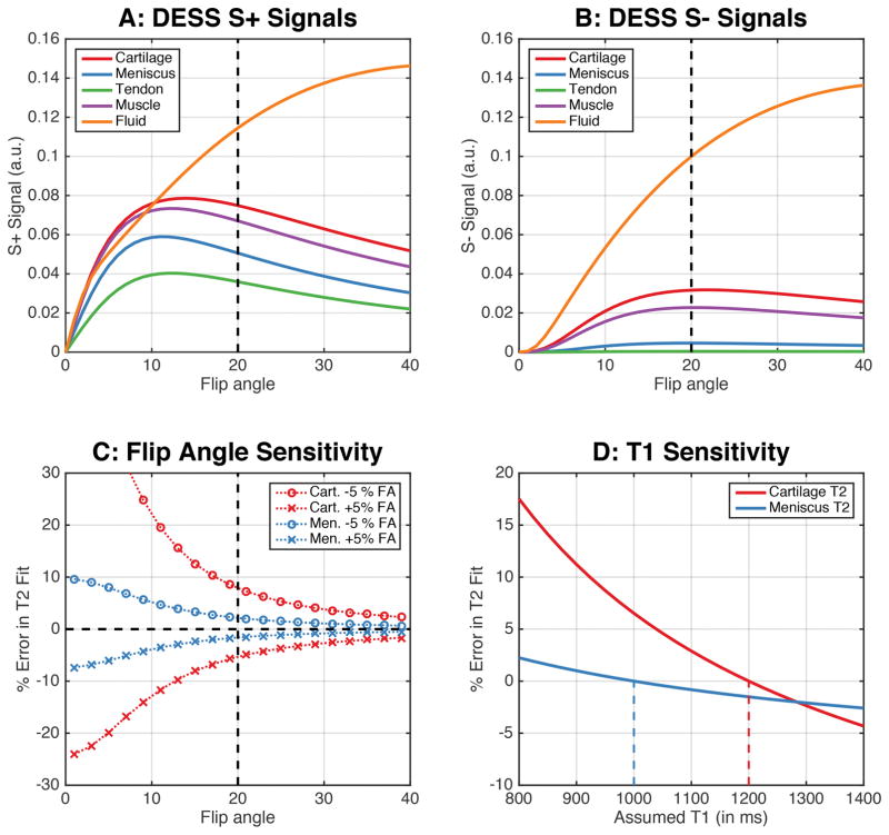
Figure 3.
Simulating the DESS sequence for different musculoskeletal tissue shows that a flip angle of 20° can maintain high signal levels in both the S+ and S− echoes (a–b) while maintaining an adequate contrast between the cartilage, meniscus, and other surrounding tissues (a). At higher flip angles, T2 measurements are less sensitive to B1 variations (c). Increasing flip angles have lower sensitivity to B1 variations and errors in T2 fits. For the T2 calculation in this study, a T1 of 1200ms and 1000ms was assumed for cartilage and meniscus (vertical dashed lines in panel D). The impact of true T1 values being different from those assumed show that ±10% T1 variations only engender <±3% variations in T2 values (d). The T1 values used for simulating the cartilage, meniscus, tendon, muscle, and synovial fluid were 1.2s, 1.0s, 1.1s, 1.4s, and 2.5s respectively while the T2 values were 40ms, 10ms, 5ms, 30ms, and 600ms, respectively.

