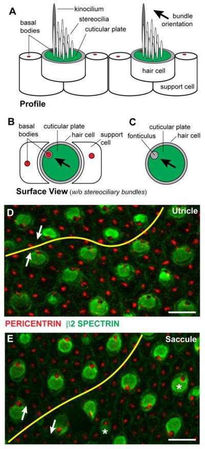
Figure 1. Vestibular hair cell PCP visualized via β2-SPECTRIN immunolabeling.

(A) Lateral profile of vestibular hair cells and supporting cells. Hair cells are readily distinguished by a polarized bundle of stereocilia projecting from the apical cell surface that is organized in a staircase fashion with the tallest stereocilia adjacent to a single tubulin based kinocilium. Stereocilia are anchored in the actin-rich cuticular plate (green) and the kinocilium is associated with a basal body (red). (B) Surface view of a vestibular hair cell without the stereociliary bundle and kinocilium illustrating the lateral position of the basal body. Hair cells are surrounded by supporting cells that also contain a single basal body however the polarized distribution of the basal bodies in these cells is not obvious. (C) In hair cells the basal body is located within a gap in the cuticular plate called the fonticulus, and the position of the fonticulus can be used as a readout of stereociliary bundle polarization and orientation. (D) β2-SPECTRIN and PERICENTRIN labeling of cuticular plates and basal bodies respectively shows the planar polarity of the utricular maculae and can be used to map the LPR (yellow line). (E) β2-SPECTRIN and PERICENTRIN labeling of the saccular maculae and the position of the LPR. The orientation of stereociliary bundles (white arrows) for pairs of cells separated by the LPR, and occasional cells that are misplaced (asters) relative to the LPR are indicated. Labeled samples were collected at P0. Scale bars are 10 μm.
