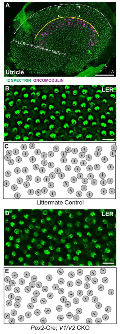
Figure 2. Vangl1 and Vangl2 are required for proper stereociliary bundle orientation in the utricular maculae.

(A) An overview of the mouse utricular maculae at P2 showing the relative positions of β2-SPECTRIN labeled hair cells analyzed in this study. The utricle was broadly divided into three domains, the Striola which contains ONCOMODULIN-expressing hair cells, and the lateral extrastriolar region (LER) and medial extrastriolar region which flank the Striola. In the utricle the approximate location of the LPR (yellow line) corresponds to the boundary between the LER and the Striola. The lateral boundary of the sensory epithelia (dashed white line) serves as a reference for stereociliary bundle orientation measures as described in the methods, and the framed region is the approximate location of images presented in B&C. (B) Vestibular hair cells from the LER of littermate control mice labeled with β2-SPECTRIN. (C) Schematic illustration of stereociliary bundle orientations for hair cells imaged in ‘B’. (D) Vestibular hair cells from the LER of Pax2-Cre; V1/V2 CKO mice labeled with β2-SPECTRIN. (E) Schematic illustration of stereociliary bundle orientations for hair cells imaged in ‘D’. Scale bars are 100μm (A) or 10μm (B,D).
