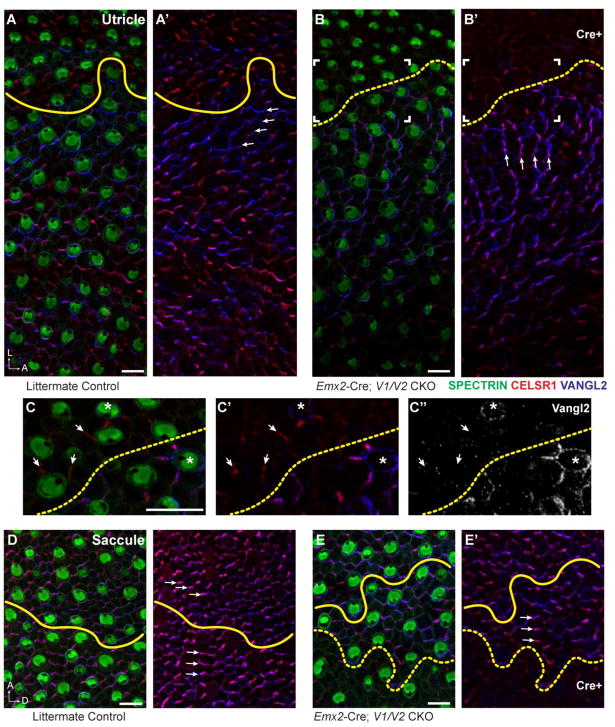
Figure 6. Domineering non-autonomy phenotype of Emx2-Cre; V1/V2 CKOs occurs in the utricular maculae.
(A,A′) Hair cell PCP, as evident in the polarized displacement of the fonticulus following β2-SPECTRIN labeling, and the asymmetric distribution of CELSR1 and VANGL2 (arrows) are aligned along a common axis that spans the LPR (yellow line) in the P0 mouse utricle. (B,B′) In Emx2-Cre; V1/V2 CKOs stereociliary bundle orientation and VANGL2 expression is disrupted in the lateral Cre-positive region of the utricular maculae. The stereociliary bundle orientation of wild type cells in the Cre-negative region are also misoriented, and adopt a new polarity axis that remains aligned with a similarly reoriented distribution of CELSR1 and VANGL2 (arrows). The framed region corresponds to panels ‘C-C” and the boundary of Cre-mediated recombination based upon VANGL2 fluorescence is indicated (dashed yellow line). (C–C″) Higher magnification image of cells at the Cre expression boundary (dashed yellow line) reveals changes in VANGL2 distribution which encircles some cells near the boundary or encircles isolated wild type cells remaining in the Emx2-Cre expressing domain (asters). CELSR1 persists at some intercellular boundaries (arrows) despite the loss of VANGL1 and VANGL2. (D,D′) In the P0 saccule hair cell PCP and the asymmetric distribution of CELSR1 and VANGL2 are similarly aligned along a common axis that spans the LPR (yellow line). (E,E′) In Emx2-Cre; V1/V2 CKOs stereociliary bundle orientation and core PCP protein distribution is not altered in wild type cells abutting the Cre-recombination boundary (dashed yellow line). Note that unlike the utricle, in the saccule the LPR and the Emx2-Cre recombination boundary are in close proximity but do not always coincide. Scale bars are 10μm.

