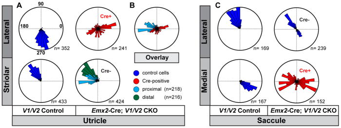
Figure 7. Quantification of domineering non-autonomy in Emx2-Cre; V1/V2 CKOs.
(A) The orientation of individual stereociliary bundles from P2 littermate controls and Emx2-Cre; V1/V2 CKO utricles graphed on circular histograms for all hair cells analyzed in the lateral and striolar regions of the utricular maculae. For controls these regions were distinguished based upon the position of the LPR while for CKOs the Cre-boundary was determined based upon remaining VANGL2 expression. (B) An overlay of histograms corresponding to a subset of wild type cells located proximal to the Cre-boundary and Cre-positive cells from the lateral region suggest that some hair cells are coordinated along a shared axis. (C) The orientation of individual stereociliary bundles for all cells analyzed in the lateral and medial regions of the saccular maculae. For controls these regions were distinguished based upon the position of the LPR while for CKOs the Cre-boundary was determined based upon remaining VANGL2 expression. Since the LPR and the Cre-boundary do not overlap in the saccule, this approach results in a bimodal distribution of bundle orientations aligned along a common axis for the Cre-negative lateral region of V1/V2 CKOs, and a unimodal distribution for the lateral region of littermate controls. For these histograms, 90° is oriented toward the lateral border of the sensory epithelia (Fig.2A,4A dashed white line) and 270° is oriented away from the lateral border. The total number of hair cells represented by each histogram (n) is shown, and bin width is 10°.

