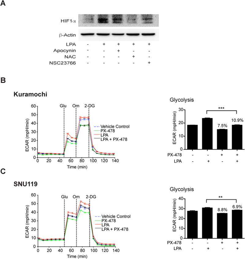
Figure 5. LPA induces pseudohypoxic increase in HIF1α via Rac1-NOX-ROS to promote aerobic glycolysis.
LPA stimulates an increase in HIF1α levels through Rac1, NOX, and ROS (Panel A). SNU119 cells, pretreated with Rac-inhibitor (NSC23766, 10 µM), NOX-inhibitor (Apocynin, 100 µM), or ROS-scavenger (N-acetyl cysteine, 10 µM) for 1 hr, were stimulated with LPA (10 µM) for 6hrs along with untreated controls. Cells were lysed and immunoblot analyses were carried out with the antibodies to HIF1α. The blot was stripped and re-probed for GAPDH to ensure equal protein loading. Results from a typical experiment (n = 3) are presented in Panel A. Role of HIF1α in LPA-stimulated aerobic glycolysis was monitored using Kuramochi (Panel B) and SNU119 (Panel C) cell lines. Cells, pretreated with HIF-1α-specific inhibitor PX-478 for 18 hours, were stimulated with LPA (10 µM) for 6 hours and ECAR analysis was carried out. ECAR flux and the rate of glycolysis were plotted (mean ± SEM; n = 9 to 12 measurements). Statistical significance was determined using Student’s t test (**P<0.005; ***P<0.0005). Percentile decreases over the basal levels of glycolysis are denoted above the bars of the chart.

