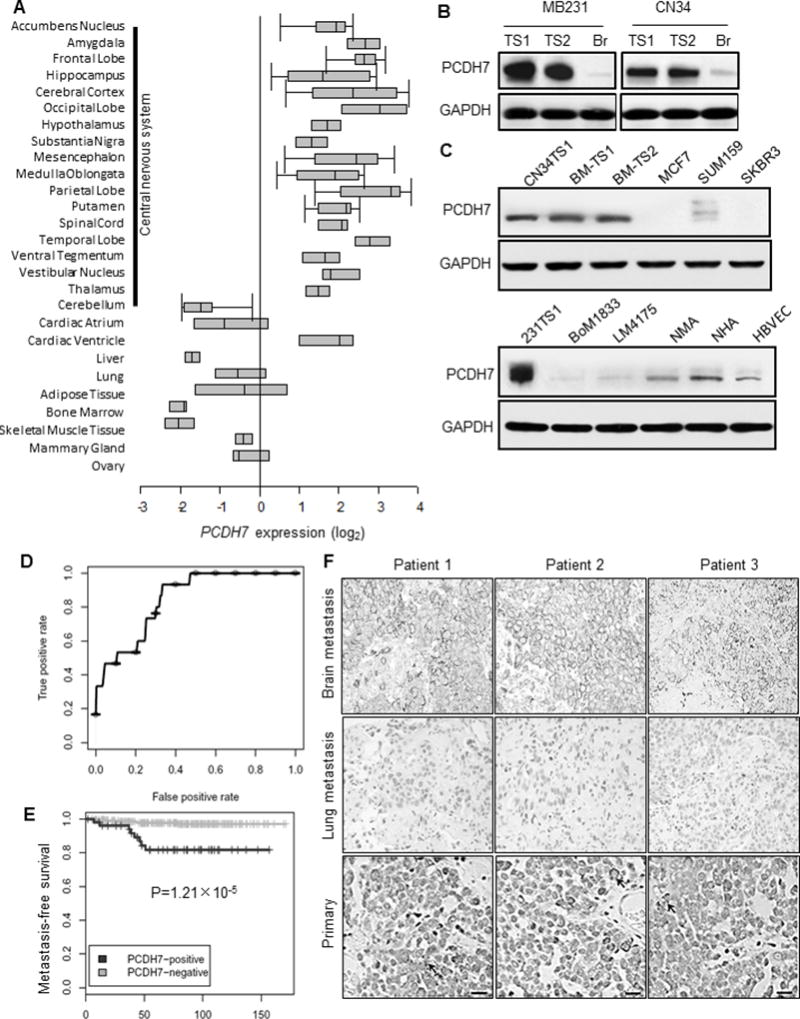
Fig. 2. High expression of PCDH7 in brain metastasis.

A. Oncomine database analysis of PCDH7 expression in normal human tissue (Neurogenetics 2006 7:67–80). B. Western Blot analysis of PCDH7 in independent brain metastasis-derived tumorspheres (TS1 and TS2) and corresponding brain-seeking (Br) cell lines. C. Protein expression of PCDH7 in TNBC patient brain metastasis tumorspheres (BM-TS1 and 2) and various cell models. MCF7, SUM159, SKBR3: human breast cancer cell lines; BoM1833: MB231 bone-seeking cell line; LM4175: MB231 lung seeking cell line; NMA: primary normal mouse astrocytes; NHA: normal human astrocytes; HBVEC: human brain microvascular endothelial cells. D. Receiver Operating Characteristic (ROC) curve for PCDH7 expression in primary breast tumor samples of brain metastatic patients using the combined 368 microarray data (MSK-82 and EMC-286 cohort). E. Kaplan–Meier curves showing the brain metastasis-free survival of patients with positive or negative PCDH7 expression in the combined cohort of 368 breast cancer patients, P=1.21×10⁻⁵ determined by log rank test. F. Representative PCDH7 immunohistochemistry staining of matched patient tissue sections of brain metastasis, lung metastasis and primary breast tumors. Scale bar: 20µm.
