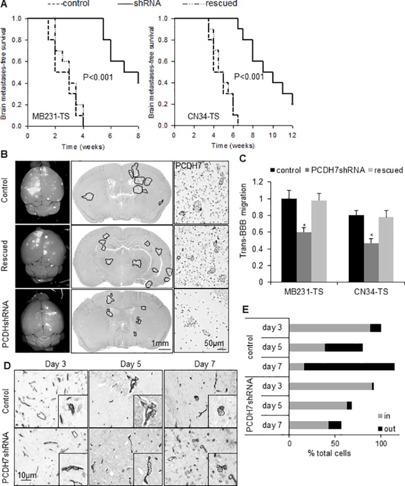
Fig. 3. Functional roles of PCDH7 in brain metastasis.

A. Kaplan-Meier curves for brain metastasis-free survival of mice injected with tumorsphere cells derived from MB231-Br (left) or CN34=Br (right) models expressing the shRNA targeting PCDH7, the control shRNA or the PCDH7 rescued. n=10 mice per group. P value was determined using log rank test. B. Representative images showing metastatic lesions in mouse whole brain (left column), H&E stained brain section (middle column), and PCDH7 immuno-reactivities in the brain metastases (right column). It’s noted that high expression of PCDH7 in both tumor cells and surrounding astrocytes in control group was diminished by PCDH7shRNA. n=5 mice per group. C. Transmigration of the indicated cells through the in vitro BBB system. * P<0.05, vs. control shRNA. D. Representative images showing intravascular and extravasated tumor cells in mouse brain sections. MB231-TS cells were visualized on 10µm-thick brain sections by anti-human CD44 and blood vessels by anti-mouse CD34 using immunohistochemistry. Black rectangles are higher magnification single cell images. CD44+ tumor cells were segmented by dash lines. E. Percentage of cancer cells located inside (in) vs. outside (out) blood vessels at indicated days after intracardiac tumor cell injection. Intravascular and extravasated tumor cells were counted in every fourth section throughout the entire mouse brain. Data was relative to “control group day 3”. It’s noted that the % total cells > 100% is due to the cell proliferation, and the % total cells < 100% is due to cell disappear. n=3 mice per each time point.
