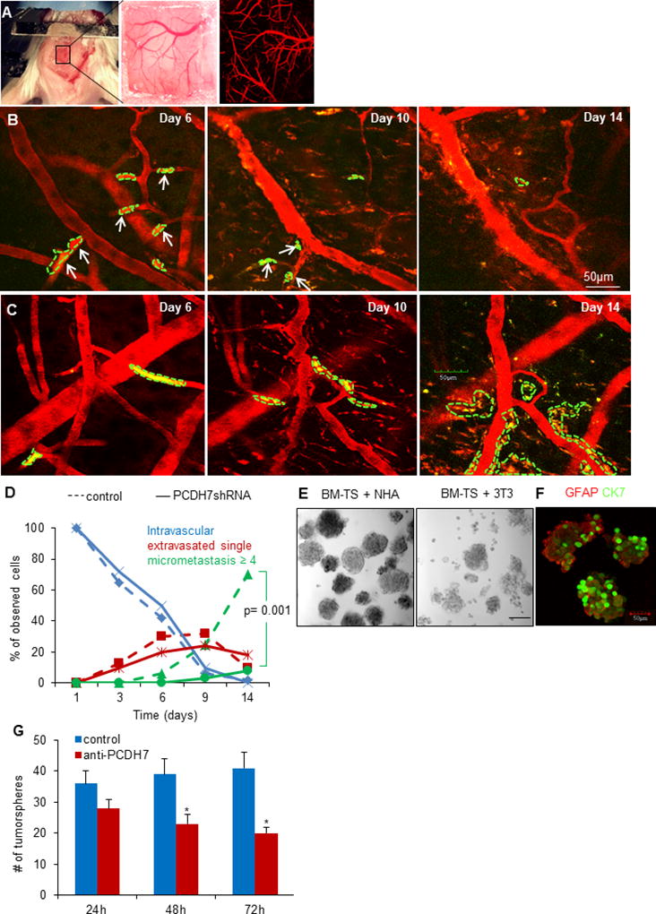
Fig. 4. PCDH7 mediates the interaction between cancer cells and astrocytes to promote tumor colonization.

A. A thinned-skull window with perfused brain vessels (Texas-red dextran, red). B. Intravital two-photon microscopy images showing tumor cell arrest and extravasation in mouse brain at day 6, and disappeared at day 10 and day 14 (indicated as solid white arrows) for PCDH7shRNA MB231-TS cells, while the control shRNA cells formed colonization at day 14 (C). It’s noted that the blood vessels became tortuous and leaky after day 6 and remodeled at day 14. D. Extravasation and colonization of the indicated cells in mouse brain over time. n=4 mice per group. E. Representative bright-filed images of tumorspheres at passage 2 when co-culturing BM-TS cells with NHA or normal fibroblasts 3T3 cells. F. Representative immunofluorescent images showing the heteotypic spheres formed by cancer cells (stained for CK7) and astrocytes (stained for GFAP). G. Effects of PCDH7 neutralization by anti-PCDH7 antibody (1:100) on tumorsphere formation (diameter >50µm) in the dissociated BM-TS cells and NHAs co-culturing system. * P<0.05, vs. control.
