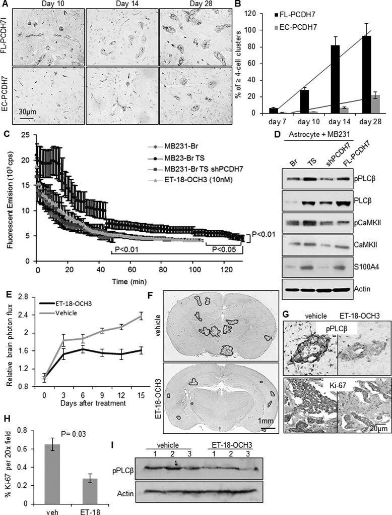
Fig. 5. Tumor-astrocyte interaction activates PCDH7-PLCβ-Ca2+ signaling to promote colonization.

A. Representative immunohistochemistry images of metastatic colonies in mouse brain sections with intra-cardiac injection of indicated cells. Tumor cells were visualized with anti-human CD44 antibody and blood vessels were visualized with anti-mouse CD34 antibody. B. Percent of ≥ 4-cell clusters located outside vessels at indicated days after tumor cell injection (n=3 mice/time point/cell line). C. Baseline level of cytoplasmic Ca2+ in the indicated tumor cells measure by ratiometric emission of calcium-saturated Asante of the tumor cells when co-culturing with astrocytes. D. Signal activity of PLCβ, CaMKII and S100A in the indicated MB231 cells when co-culturing with normal human astrocytes. E. In vivo effects of PLC selective inhibitor ET-18-OCH3 (i.p., 30mg/kg, once daily) on brain metastatic tumor growth. n=10 per group. P=0.04, determined by ANOVA post-hoc test. F. Representative whole-brain sections showing macro-metastatic lesions of ET-18-OCH3 or vehicle-treated mice at the end of the treatment. G. Representative immunostained images of pPLCβ1 and Ki-67 in brain sections. H. Quantification of the % of Ki-67-positive tumor cells in the brain sections. P=0.03, determined by student’s t-test. I. Western blot of pPLCβ1 expression in mouse brain lysates. N=3 per group.
