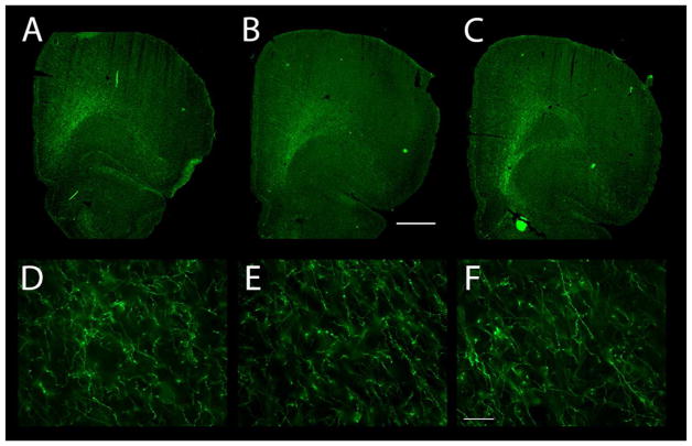
Figure 10.
Dopaminergic innervation of the OFC as revealed by TH immunoreactivity. (A–C) The distribution of TH-ir axons is shown at three different levels, from rostral (A) to caudal (C). Note the sweeping band of TH-ir axons in the ventrolateral frontal cortex, running diagonally from the white matter to the pial surface. The dopamine innervation completely fills AId2 and extends somewhat medially (into LO) and laterally (into AId1). (D–F) TH-ir axons in Layer 6 (panel D), Layer 5 (E), and Layer 3 (F). Scale bars: A–C, 1000μm and D–F, 50μm.

