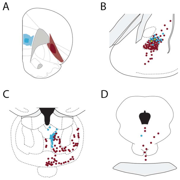
Figure 12.
Localization of retrogradely-labeled cells in the (B) mediodorsal thalamus, (C) basolateral amygdala, and (D) midbrain (including rostral dorsal raphe) after (A) depositing Fluoro-Gold into the mPFC (blue) and cholera toxin B into the OFC (red). The filled blue circles indicate neurons retrogradely-labeled from the mPFC and red filled circles indicate cells retrogradely-labeled from the OFC; yellow stars depict double-labeled cells. A single midbrain cell and two amygdala cells (yellow stars) were double labeled.

