
Figure 5.
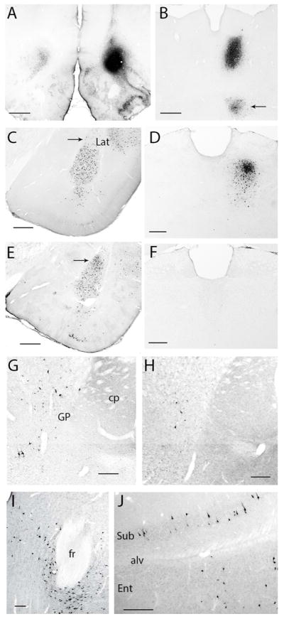
Retrograde labeling following tracer deposits into the OFC. (A) Tracer deposit into VLOp and contralateral labeling (case A1062). (B) Dense retrograde labeling in case A1052 in the central segment of MD, with the arrow pointing to labeling in the submedius nucleus. (C, E) Retrograde labeling in the amygdala following tracer deposits in AId2 (C; A1014) and the LO (E; A1064). Note the greater density of labeled cells in the lateral nucleus after FG was iontophoresed into the LO. (D, F) Retrograde labeling in the MD following tracer deposits into either the LO (D; A1065 or the DI-GI (F; A1060). (G, H) Retrogradely-labeled cells in the globus pallidus and substantia innominata following a deposit into AId (G; A1016) or DI-GI (H; A1061). Labeled neurons in the parafascicular nucleus (I) and subiculum and entorhinal cortex (J) in case A1014 (AId2 deposit). Scale bars: (A) 1000 μm; (B, C, E) 500 μm; (D, F–H, J) 250 μm; (I) 250 μm. Abbreviations: alv, alveus; CP, caudatoputamen (striatum); fr, fasciculus retroflexus; ENT, entorhinal cortex; GP, globus pallidus; Lat, lateral amygdala nucleus; PF, parafascicular nucleus; Sub, subiculum.