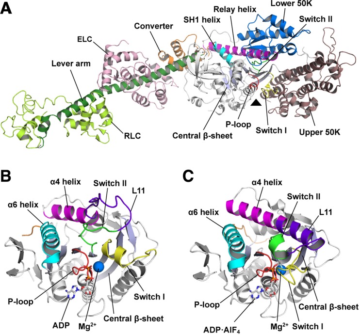
Fig. 1.
Structures of myosin and kinesin. a Ribbon diagram of the motor domain, lever arm, essential light chain (ELC) and the regulatory light chain (RLC) of myosin II (scallop myosin S1; PDB 1SR6). The triangle indicates the nucleotide-binding site. b The motor domain of ADP-bound kinesin (KIF5B; PDB 1BG2). ADP and Mg2+ are represented by stick and sphere models, respectively. c The motor domain of ATP-bound KIF5B (PDB 4HNA). ADP·AlF4 (an ATP analogue) and Mg2+ are represented by stick and sphere models, respectively. The individual architectures related to motor function are shown with different colours in each panel

