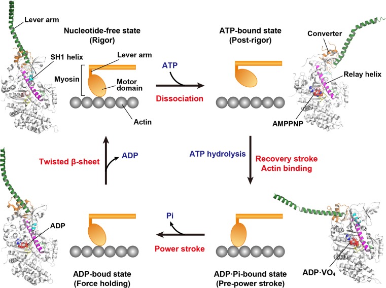
Fig. 2.
Mechanochemical cycle of myosin. The structural models with ribbon diagrams are created by the crystal structures of myosin II (scallop myosin S1; upper left, PDB 1SR6; upper right, PDB 1KQM; lower right, 1DFL; lower left, 3I5F). The lever arm (dark green), converter subdomain (orange) and SH1 helix (cyan) adopt different orientations and conformations in each state. The nucleotide-binding regions, including the P-loop, switch I and switch II, are coloured in red, yellow and green, respectively. Each nucleotide in the motor domain is represented by a sphere model. A power stroke corresponds to a step of force generation

