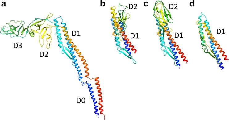
Fig. 3.
Structural comparison of flagellin from various bacteria. a Cryo-electron microscopy (Cryo-EM) structure of full-length flagellar filament protein, flagellin (FliC), from Salmonella (PDB ID 1ucu). b–d Crystal structures of Burkholderia pseudomallei FliC (BpFliC; PDB ID 4cfi) (b), Pseudomonas aeruginosa FliC (PaFliC; PDB ID 4nx9) (c), and Bacillus subtilis FliC (BsFliC; (PDB ID 5gy2) (d). The D0 domains of Bp-, Pa and Bs-FliC were removed for crystallization. The chains are colored in the rainbow sequence of colors from blue to red for the N- to C-terminus

