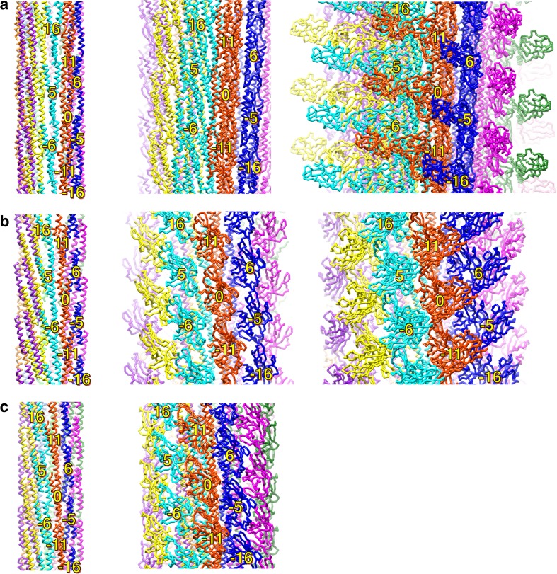
Fig. 5.
Intersubunit domain interactions in the axial structure. Side views of the R-type filament (a), the hook (b), and the rod (c) showing the subunit arrangement and packing of the different layers: left panel shows the array of the D0 domains; middle panel shows the D1 domains; right panel shows the D2 and D3 domains. Individual protofilaments are shown in different colors. The subunits surrounding subunit 0 are labeled with the number showing the direction of the helical line

