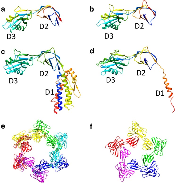
Fig. 8.
Crystal structures of the capping protein of the bacterial flagellar filament (FliD) from various bacteria. Ribbon drawings of StFliD (PDB ID 5h5t) (a), Serratia marcescens FliD (SmFliD; PDB ID 5xlk) (b), Escherichia coli FliD (EcFliD; PDB ID 5h5v) (b), and PaFliD (PDB ID 5fhy) (d) are shown in rainbow sequence of colors from blue to red for the N- to C-terminus. e, f Structure of the FliD cap assembly: e EcFliD hexamer (PDB ID 5h5v), f StFliD pentamer (PDB ID 5h5t). Subunits are represented in different colors

