
Fig. 2.
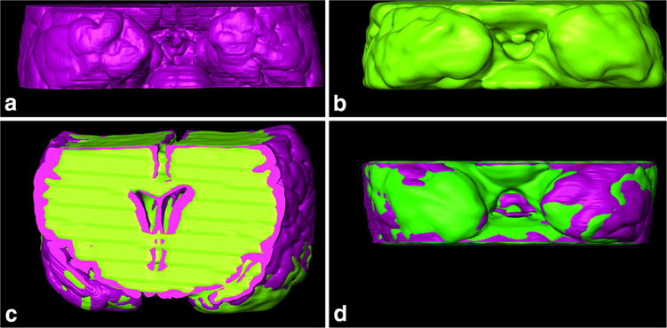
Histology-to-MRI manual 3D registration of Case 1. a Basal central block surface segmented from histological slices (purple); b basal central block surface segmented from T1 MRI images (green); c frontal view of 3D registered histology and MRI, and d basal view of 3D registered histology and MRI. These outlines were used to calculate DSC and nWSD 3D values