
Fig. 4.
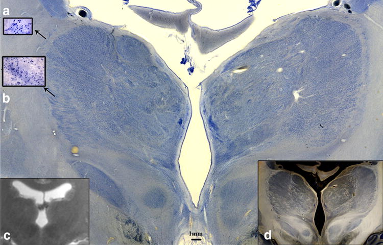
Coronal Nissl-stained 560 lm section (Case 3) at the level of the thalamus and subthalamic nuclei (STN) a High power magnification view of the reticular nucleus and b lateral group. c 3.0 T MRI of the same specimen, d Dark-field illumination of the same slice, showing contrast enhancement and myeloarchitecture as criterium for segmentation