
Fig. 5.
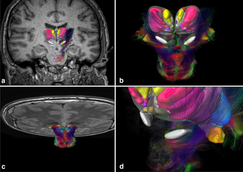
Registered MRI and DTI with cytoarchitectonic fields and subthalamic region. a Coronal MRI fused to histology. b Fiber tracking of brainstem fused to 3D histology of STN and thalamus. c Horizontal MRI fused to DTI and histology. d Detail of the subthalamic region and fibers surrounding the histology-defined STN