
Fig. 8.
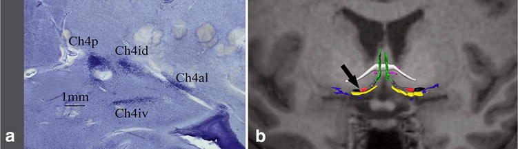
a Microscopic view of basal forebrain components in a coronal slice at the level of the black arrow in b; b basal forebrain cholinergic system (BFCS) histological map fused to T1 MRI of the same subject. White: anterior commissure; green: CH2; yellow: CH3; gray: CH4am; red: CH4i, black: CH4p; blue: Ayala’s nucleus; pink: nucleus juxta commissuralis. Color coding follows Grinberg and Heinsen (2007), Mesulam’s nomenclature is adapted from Mesulam et al. (1983)