
Figure 2.
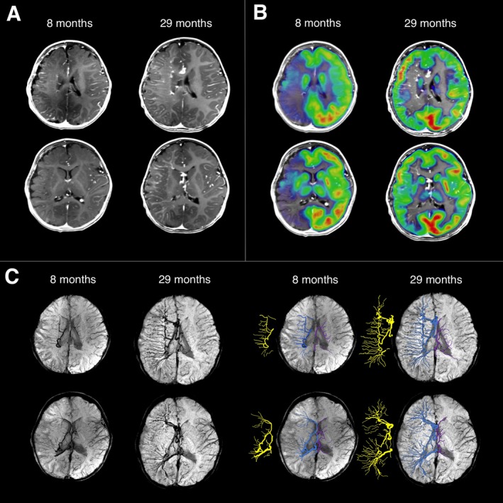
Multimodal neuroimaging at age 8 months and 29 months. (A) Postcontrast T1‐weighted images showing leptomeningeal enhancement and lack of normal surface veins in the right frontal, parietal, and temporal lobes at both time points. Moreover, the right hemispheric multilobar atrophy at age 29 months seemed to be slightly less pronounced than at age 8 months, with less asymmetric CSF space. (B) On interictal 2‐deoxy‐2‐[18F]fluoro‐D‐glucose (FDG)‐PET scans (coregistered with postcontrast T1 images), severe right hemispheric hypometabolism was observed, with a relatively preserved medial frontal and parietal cortex at the age of 8 months. Glucose metabolism at the age of 29 months showed a dramatic improvement of metabolic activity. (C) Susceptibility‐weighted imaging (SWI) minimum intensity projection (MIP) images visualized enlarged deep medullary veins in the right hemisphere (enhanced in blue on the right panels, also its skeleton is displayed next to the enhanced images in yellow; contralateral deep medullary veins are enhanced in purple). The right‐sided deep veins became much more extensive by 29 months of age, encompassing the whole right hemisphere.