Fig. 3.
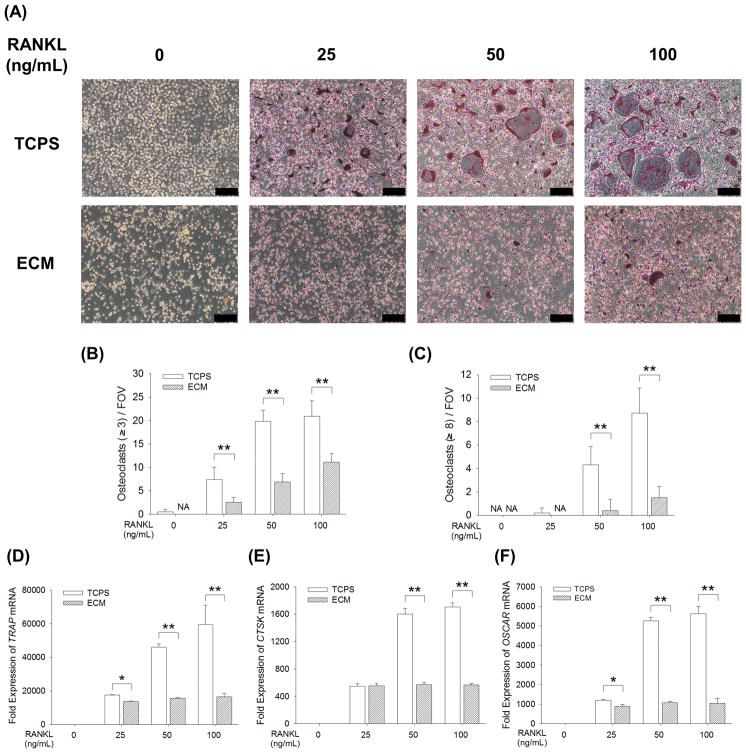
Cell-derived ECM inhibited in vitro osteoclastogenesis of BMMs. BMMs were cultured on the standard TCPS or ECM-coated substrate and induced toward osteoclasts by 25 ng/mL, 50 ng/mL, or 100 ng/mL RANKL, supplemented with 20 ng/mL M-CSF. (A) After a five-day culture, multinucleated osteoclasts were positively stained for TRAP and the nuclei were counterstained with DAPI (three replicates for each treatment). Scale bar = 200 μm. (B–C) Quantification of the cell number of TRAP-positive osteoclasts with greater than 3 nuclei (B) and with 8 or more nuclei (C). TRAP-positive osteoclasts were counted in at least ten randomly chosen fields of view (FOV). (D–F) The mRNA levels of TRAP (D), CTSK (E), and OSCAR (F) were measured by real-time RT-PCR. Values are presented as the mean ± S.E.M. of four independent experiments (n = 4). Statistically significant differences are indicated by * p < 0.05 or ** p < 0.01 between the indicated groups.

