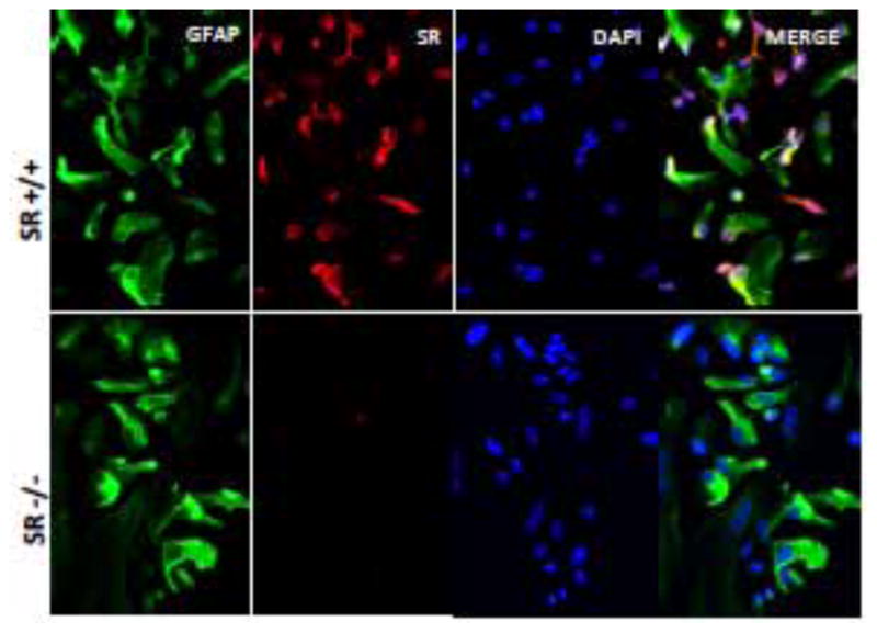
Figure 1. Immunocytochemical demonstration of glial fibrillary acid protein and serine racemase in astrocytes after seven days of culture.

GFAP is green; SR is red; and the nuclei are stained blue with DAPI. Note the absence of SR in cultures from SR−/− mice.
