
Figure 7. ERK regulates FOXC2 activity and DLL4 expression in mouse lung EC in vivo .
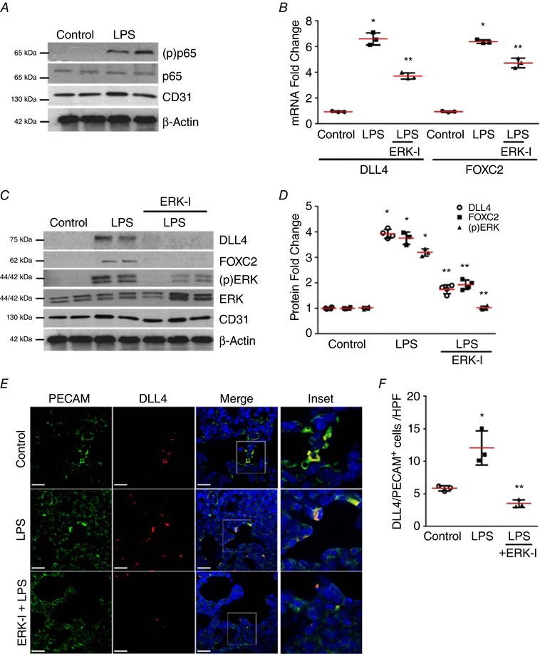
A–D, mouse lung EC isolated from 4‐day old mice 24 h after LPS treatment with or without i.p. 20 mg kg−1 ERK‐I were used. A, phosphorylation of the NF‐κB sub‐unit p65 was assessed in lung EC by Western blotting using an anti‐(p) p65 antibody, n ≥ 4 mice in each group. B, lung EC RNA was used to quantify DLL4 and FOXC2 mRNA by qRT‐PCR, P < 0.01 (*Control vs. LPS: ** LPS vs. ERK‐I + LPS), n = 3. C, DLL4, FOXC2, and (p)ERK were examined in lung EC by Western blotting, and quantified by densitometry (D), P < 0.01 (*Control vs. LPS; **LPS vs. ERK‐I + LPS), n ≥ 3. E and F, mouse lung paraffin sections obtained after LPS and ERK‐I treatments were used for studies. E, confocal microscopy depicting PECAM (green), DLL4 (red), and DAPI (nucleus‐blue) immunofluorescent images. Images were captured at 63×, with the inset being the magnification of the boxed area in the Merge image. F, graph summarizing percentage of PECAM+/DLL4+ cells per high power field (HPF), P < 0.01 (*Control vs. LPS; ** LPS vs. ERK‐I + LPS), n ≥ 3 mice. Scale bar represents 20 μm.