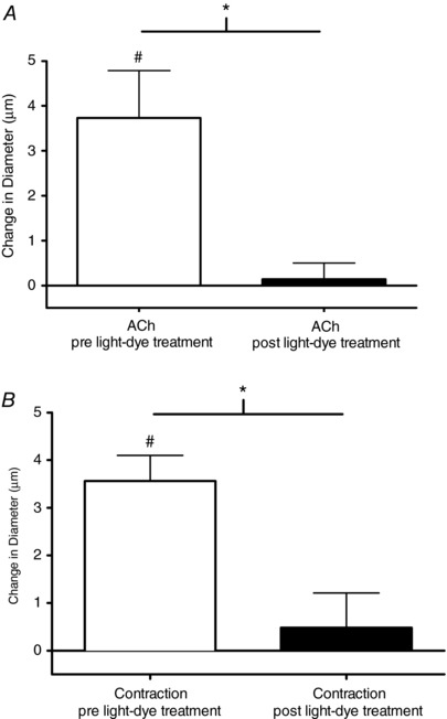
Figure 2. Ablation of cells between the capillary stimulation site and the upstream 4A arteriolar observation site eliminated upstream 4A arteriolar vasodilatation.

The change in diameter in response to capillary stimulation using micropipette application of ACh (A) or microelectrode stimulation of a bundle of skeletal muscle fibres (B) under control conditions (white bars) and after ablation of cells between the stimulation site and the 4A arteriolar observation site (black bars). #Vasodilatation significantly different from 0. *Significant differences between conditions.
