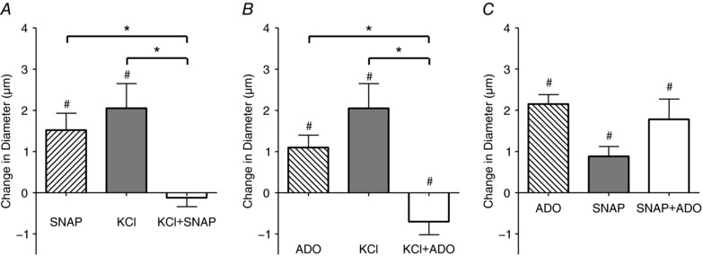
Figure 4. Simultaneous addition of vasodilators demonstrates inhibition between vasodilators.

The change in diameter in of the 4A arteriole in the last minute of drug application in response to 10−7 m SNAP alone (□), 10 mm KCl alone (△) and 10−6 m SNAP+10 mm KCl added together simultaneously (■) (A); 10−6 m ADO alone (□), 10 mm KCl alone (△) and 10−6 m ADO+10 mm KCl added together simultaneously (■) (B); 10−6 m ADO alone (□),10−6 m SNAP alone (△) and 10−6 m ADO+10−6 m SNAP added together simultaneously (■) (C). #Vasodilatation is significantly different from 0. *Significant differences between conditions. Protocol details can be found under Protocol 2 and raw baseline and maximal diameters can be found in Table 2.
