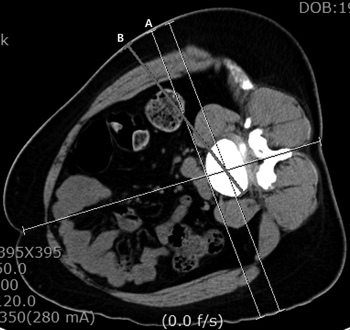
Figure 2.

Preoperative planning for the location of the skin incision. Axial view on computed tomography (CT) scan of an L4–L5 level with the patient in the right lateral decubitus position. Line A tangentially goes through the anterior wall of the vertebral body. Line B illustrates the ideal vector for which the cage would be placed in the intervertebral space to realize the orthogonal maneuver further.
