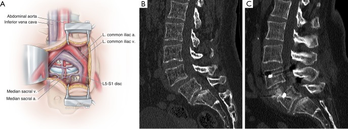
Figure 4.
Anterior lumbar interbody fusion (ALIF) and reduction of spondylolisthesis. (A) Illustration of a mini-open ALIF approach, in which the peritoneum is reflected laterally to expose the retroperitoneal vessels and spine. At L5-S1, the working corridor is typically between the iliac vessels below their bifurcation. This allows a full anterior-posterior exposure of the disc space in its entirety. At L4–5 and higher lumbar levels, the approach is limited by how much mobilization of the vascular structures can be achieved. a., artery; L. left; v., vein; (B) preoperative sagittal computed tomogram (CT) of a 53-year-old woman with intractable back pain demonstrates a grade I spondylolisthesis at L4–5 that was demonstrated to be mobilized on flexion-extension imaging; (C) postoperative CT of the same patient after a minimally invasive L4–5 ALIF and placement of posterior percutaneous pedicle screws demonstrates full reduction of the spondylolisthesis and clinically significant restoration of disc height. Used with permission from Barrow Neurological Institute, Phoenix, Arizona.

