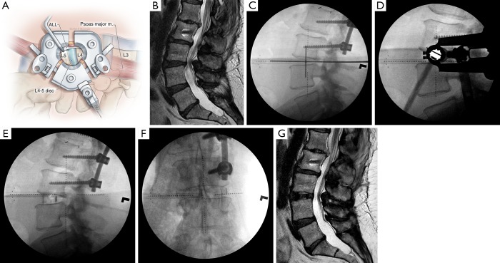
Figure 5.
Overview of lateral lumbar interbody fusion (LLIF). (A) Illustration of the LLIF operative field through a minimally invasive retractor allows visualization of most of the lateral length of the disc space, as well as the anterior longitudinal ligament (ALL), which typically is kept intact except when anterior column realignment is necessary. m., muscle; (B) preoperative sagittal T2-weighted magnetic resonance image (MRI) of a 57-year-old woman with a previous L1-L3 fusion and distal adjacent segment instability at L3-4, with resultant severe central canal stenosis causing intractable neurogenic claudication; (C) intraoperative lateral fluoroscopy with a cross hair is used to mark the incision on the patient’s right flank. The center of the incision is approximately one-third of the anterior-posterior (AP) length of the disc space. At L4–5, it is centered on one-half of the AP length; (D) intraoperative lateral fluoroscopy shows the docking location of the minimally invasive lateral retractor; (E) lateral and (F) AP radiographs taken immediately after placement of an LLIF interbody cage demonstrate distraction of the disc space and increased segmental lordosis. A long interbody is placed, spanning the entire wide of the disc space from each diaphysis; (G) postoperative MRI demonstrates restoration of the L3–4-disc height and indirect decompression of the central canal. Used with permission from Barrow Neurological Institute, Phoenix, Arizona.

