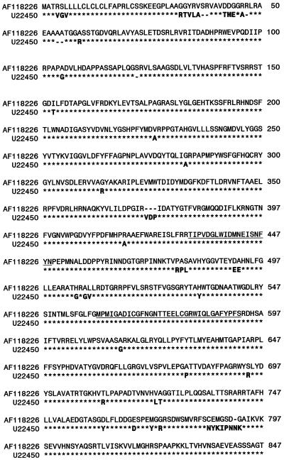
Figure 6.
Alignment of deduced protein sequences from the high-pI α-glucosidase genomic clone (AF118226, cv Igri) and cDNA clone (GenBank U22450, cv Morex). Identical residues are marked with asterisks (*). Conserved signature regions I and II in glucoside hydrolase family 31 are underlined (Frandsen and Svensson, 1998).

