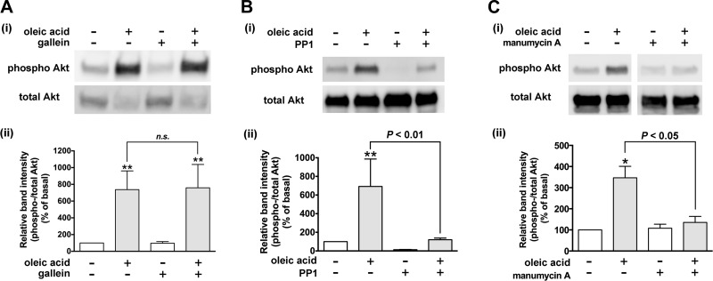
Fig. 8.
Effects of inhibitors of Gβγ subunits, ras, or Src on oleic acid-induced phosphorylation of Akt in HASM cells. Cells were pretreated with the Gβγ signaling inhibitor gallein (10 µM for 60 min; n = 7) (A), Src inhibitor PP1 (10 µM for 60 min; n = 8) (B), or ras inhibitor manumycin A (C) (3 µM for 3 h; n = 3) before treatment with oleic acid (10 µM) for 10 min. The cell lysates were processed to detect phosphorylated (Ai–Ci, top) and total (Ai–Ci, bottom) levels of Akt by immunoblot. Aii–Cii: graphical analysis. Phosphorylation of Akt is presented a ratio of phosphorylated to total Akt and expressed relative to basal ratios. Data represent means ± SE. *P < 0.05; **P < 0.01, compared with basal. White vertical spaces between the lanes in C indicate that these lanes were located on the same immunoblot but were not located in neighboring lanes on the original gel and immunoblot image.

