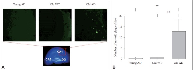
Figure 5.
Amyloid plaques in long-term OHSC from old 3xTg-AD mouse. A: Amyloid plaques (bright green), stained with thioflavin-S, were exclusively shown in the 4-week-cultured hippocampal slices from old 3xTg-AD mouse. Photos were taken in the CA1. B: The number of amyloid plaques were counted bya blind experimenter in the whole hippocampus. Each bar, mean±SE. Scale bars, 100 μm. **p<0.01 by Bonferroni post hoc analysis. AD: Alzheimer’s disease (model), WT: wild type, CA: cornus ammonis, DG: dentate gyrus.

