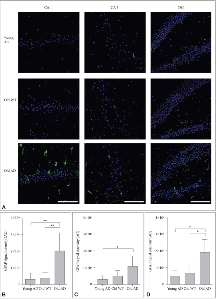
Figure 6.

Activated astrocytes in long-term OHSC from old 3xTg-AD mouse. A: Representative confocal images show astrocytes stained with anti-GFAP-antibody (green) and DAPI-stained nuclei (blue) in the 4-week-cultured hippocampal slices from young 3xTg-AD, old WT C57BL/6J and old 3xTg-AD mice. GFAP signal intensity was quantitatively measured in (B) CA1, (C) CA3, and (D) DG regions. Each point, mean±SE. Scale bars, 100 μm. *p<0.05, **p<0.01 by Bonferroni post hoc analysis. AD: Alzheimer’s disease (model), GFAP: glial fibrillary acidic protein, DAPI: 4’,6-diamidino-2-phenylindole, WT: wild type, CA: cornus ammonis, DG: dentate gyrus.
