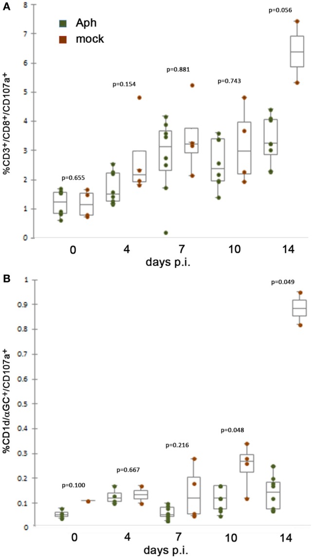
Figure 1.

At 10 days or later p.i., in vivo splenic CD8 and NKT cytotoxic lymphocyte CD107a expression is lower after infection by A. phagocytophilum in HL-60 cells compared to mock infection by uninfected HL-60 cells. (A) Splenic CD8 T lymphocytes from mock-infected (uninfected HL-60 cells) animals displayed mobilized cytotoxic responses as measured by surface CD107a expression at higher levels than with A. phagocytophilum-infected HL-60 at 14 days p.i. (B) Splenic NKT cells from infected animals demonstrate similar lack of significant cytotoxicity as measured by expression of CD107a compared to animals mock infected (uninfected HL-60 cells) at days 10 and 14 p.i. Individual points represent values from individual animals; box and whisker plots show median, first and second quartiles, and maximum and minimum values for each group. p Values are displayed comparing groups on individual days. Aph, Anaplasma phagocytophilum.
