
Figure 3.
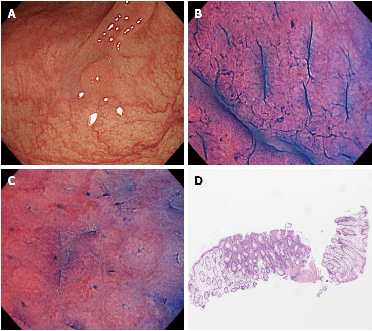
A case of disagreement between endocytoscopic (EC) 1b diagnosis and histopathologic diagnosis (adenoma). A: Flat-type lesion (0-IIa) 3 mm in size; B: The polyp was located in the sigmoid colon. The EC images showed slit-like smooth lumina; C: The EC images showed roundish lumina; D: The nuclei were not clear due to inadequate staining. An endoscopist diagnosed the polyp as EC1b. Histologic examination revealed an adenomatous polyp.