
Figure 4.
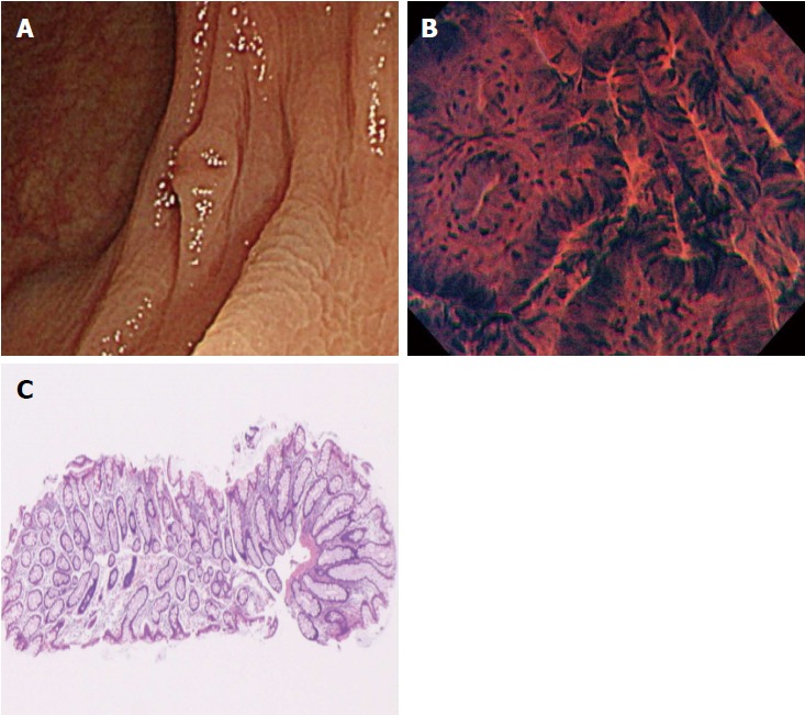
A case of disagreement between endocytoscopic (EC) 2 diagnosis and histopathologic diagnosis (hyperplastic polyp). A: Flat-type lesion (0-IIa) with a shallow depressed area, 3 mm in size; B: The polyp was located in the transverse colon. Endoscopic imaging in the shallow depressed area showed typical EC2 features with slit-like smooth lumina and uniform fusiform and roundish nuclei; C: The polyp was diagnosed as EC2. Histologic examination revealed a hyperplastic polyp without a depressed area.