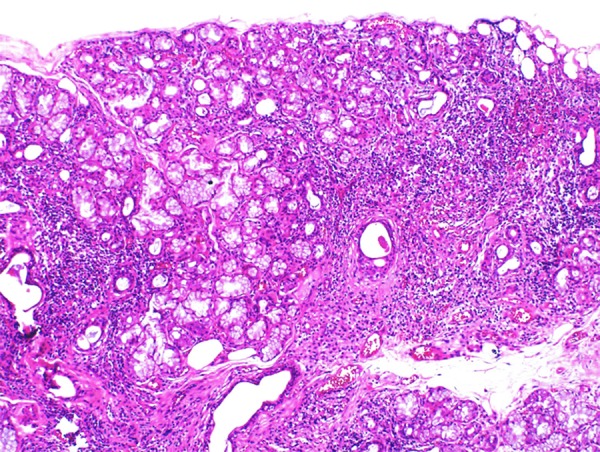
Figure 2.

Photomicrograph of the histology of the biopsy of the minor salivary gland. Histology of the biopsy of the minor salivary gland shows a diffuse mononuclear inflammatory lymphoplasmacytic infiltrate with the formation of groups of >50 lymphocytes equivalent to Chisholm-Mason stage IV (based on assessing a 4 mm2 area of salivary gland tissue) and Greenspan focus score (FS) of 2.
