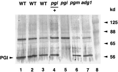
Figure 5.
Immunoblot analysis of the plastidial PGI protein in wild-type (WT) and mutant leaf extracts. Leaf extracts from the wild type (containing 24, 18, and 12 μg of proteins in lanes 1–3, respectively), pgi1/+ (24 μg of proteins in lane 4), pgi1, pgm1, adg1 (24 μg of proteins in lanes 5–7), and prestained molecular mass markers (lane 8) were separated in a 10% (w/v) SDS PAGE, electroblotted onto a nylon membrane, and probed with the antiserum against Arabidopsis plastidial PGI protein. Bands of the expected sizes were detected by the antiserum, as indicated. There were cross-reactive bands detected by the antibodies. These may be related proteins sharing antigenic sites with the Arabidopsis plastidial PGI protein or E. coli-derived proteins.

