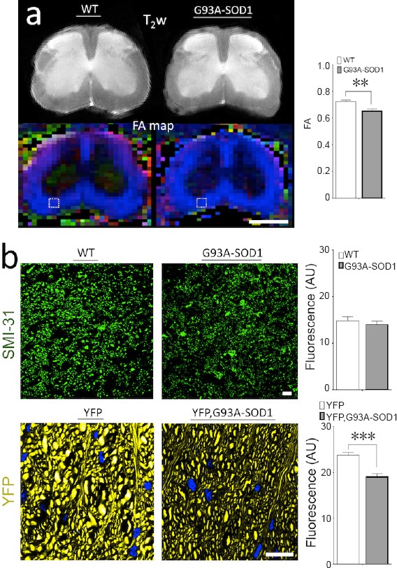
Figure 1.

Diffusion tensor imaging (DTI) can detect early microstructural changes in white matter (WM) organization in spinal cord (SC) of the SOD1 mice.
(a) High-field MR diffusion T2-weighted (T2w) imaging and in vivo fractional anisotropy (FA) maps from axial lumbar sections in control mice (WT) and ALS mice (G93A-SOD1). Analysis of regions of interest (ROIs) were performed in the anterolateral funiculi (ALF) of the ventral SC WM based on previous ex vivo studies. Comparison between both groups showed a significant early decrease (P < 0.001) of FA (index of axonal organization) in the disease group. These results prove that presymptomatic anomalies in WM microstructure can be detected in vivo by DTI. (b) Microscopy analysis using immunohistochemistry (IHC) techniques from comparative WM ROIs using a phosphorylated neurofilament marker (SMI-31) at early stages of the disease demonstrated relative signs of WM structural anomalies in the ALS mice group. However, quantitative IHC methods, did not show significant differences of this marker in the ALS mice. To improve axonal visualization, we bred a line of mice expressing endogenous yellow fluorescence protein (YFP mouse) with ALS mice creating a fluorescent mice reporter (YFP, G93A-SOD1 mice). Confocal fluorescence microscopy at a higher magnification shows alteration in size and morphology of individual axons. Further ALF quantitative confocal fluorescence analysis demonstrated a significant reduction of the fluorescence levels in the YFP, G93A-SOD1 mice. Nuclear counterstaining with DAPI (blue). Statistical analysis performed with unpaired t-test analysis (n = 5 animals per group). **P < 0.01, ***P < 0.001. Scale bars: 1 cm in a, 10 μm in b. This review was partially based on previous work from Gatto et al. (2018).
