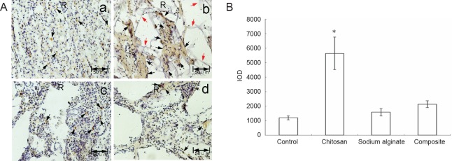
Figure 7.
NF-H expression at the spinal cord injury site of experimental rats sixty days after surgery.
(A) Immunohistochemical staining of NF-H protein; no scaffold (a), chitosan (b), sodium alginate (c), or chitosan-sodium alginate composite (d) scaffolds were implanted into the injury site of the rats. Sixty days after surgery, the spinal cords were excised, fixed, embedded in paraffin, stained with anti-NF-H antibody and restained with hematoxylin. The sections were photographed under a dissecting microscope. Brown represents NF-H immunoreactivity and purple represents nuclei. Black arrows point to NF-H immunoreactivity. Red arrows point to undegraded chitosan scaffold. Scale bars: 50 μm. (B) Quantitative analysis of NF-H expression (mean ± SD, n = 9, one-way analysis of variance followed by the Student-Newman-Keuls post hoc test; *P < 0.05, vs. control group). Control: Control group; chitosan: chitosan scaffold group; sodium alginate: sodium alginate scaffold group; composite: composite material scaffold group. NF-H: Neurofilament-H; IOD: integrated optical density.

