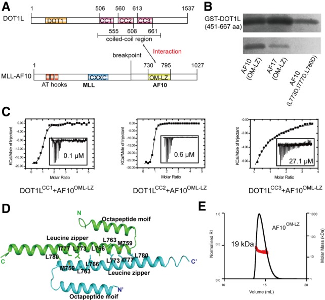
Figure 1.

The coiled-coil region of DOT1L binds to AF10 and AF17. (A) Schematic diagrams of the domain organization of DOT1L and MLL-AF10. Interactions between DOT1L and AF10 are indicated by a two-way arrow. There are three coiled-coil regions in DOT1L. (B) The coiled-coil region of DOT1L robustly binds to AF10 and AF17 by GST pull-down assay. Triple mutation AF10L773D, I777D, L780D abolishes the DOT1L binding. (C) ITC-based measurements quantifying the binding affinities between the coiled-coil region of DOT1L and AF10OM-LZ. (NB) No binding. (D) Cartoon representation of the AF10OM-LZ structure. The two chains are colored cyan and green, respectively. The residues involved in the dimer interface are shown in stick representations. (E) Size exclusion chromatography coupled with multiangle light-scattering analysis of AF10OM-LZ.
