
Figure 3.
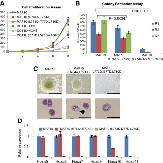
The DOT1L–AF10 binding is essential for MLL-AF10-associated leukemogenesis. (A) Cell proliferation assay for MLL-AF10 (MAF10) wild-type or mutant transduced bone marrow cells as well as MAF10 wild type transduced with or without DOT1L deletion bone marrow cells with DOT1L wild-type or mutant overexpression. Error bars indicate standard deviation (SD) from duplicates. The results were repeated at least three times. (B) Myeloid colony formation assay for transduced bone marrow cells with MAF10 wild type or mutants as indicated. Colony counts were summarized from primary, secondary, and tertiary plating on methycellulose medium in the presence of IL3, IL6, stem cell factor (SCF), and granulocyte-macrophage colony-stimulating factor (GM-CSF). Error bars indicate SD from duplicates. The results were repeated at least three times. P < 0.0001, two-way ANOVA test. (C) Representative colonies (top) and Wright-Giemas-stained cells (bottom) from the tertiary plating of MAF10 wild type or mutant transduced bone marrow cells are shown. Bars: colonies, 200 µm; Wright-Giemas-stained cells, 50 µm. (D) Real-time PCR for HOXA genes in cells as indicated. Gene expression was normalized against GAPDH and is presented as fold change against their respective levels in MAF10 wild-type cells, which was arbitrarily set at 1. Means and SDs (as error bars) from at least three independent experiments are presented.