
Figure 1.
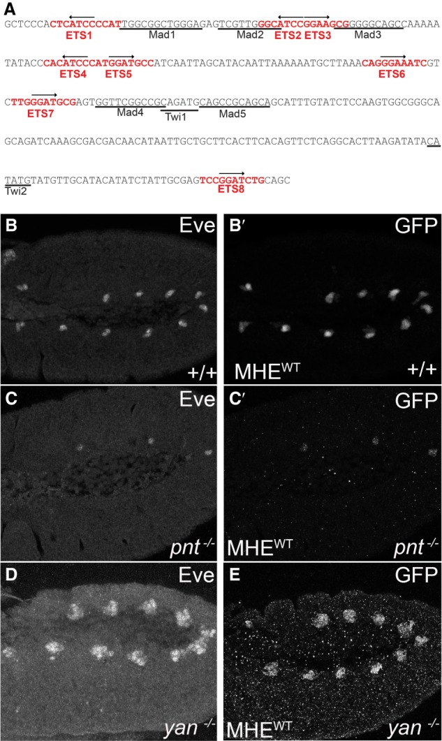
The MHE reliably reports the pattern of mesodermal Eve expression. (A) MHE sequence with putative ETS sites in red. Black arrows indicate site orientation. Previously characterized Mad and Twi sites are underlined. (B–E) Lateral views, oriented with anterior to the left and ventral down, of the thoracic and abdominal segments of representative stage 11 embryos expressing two copies of the MHEWT-GFP reporter in w1118 (B), pntΔ88 (C), or yanER443 mutants (D,E). Costaining with anti-Eve (B,C) and anti-GFP (B′,C′) shows that the reporter-driven pattern matches closely that of endogenous Eve. (D,E) Loss of yan expands these Eve expressing and GFP-expressing cell clusters.